This article reveals the differences between atelectasis vs pneumothorax (collapsed lung):
Atelectasis
It is the collapse of a segment or lobe of the lung or an entire lung. Surgery is one of the common causes of the condition, particularly the one involving your chest or upper abdomen.
Atelectasis of the lung after inhalation anesthesia in surgical patients in the United States occurs in about 87 percent of cases, according to the American Journal of Respiratory and Critical Care Medicine.
Symptoms
There may be no obvious symptoms of atelectasis. But, if you do experience symptoms, they may include:
- paleness of the skin can occur in case a large area of the lung is affected;
- coughing (mild);
- decreased expansion of the chest wall;
- stabbing pain in the chest;
- increased temperature;
- due to the transportation of oxygen being affected, your heart rate can be increased;
- cyanosis (a bluish cast to the skin and mucous membranes), especially in people who are suffering from chronic forms;
- accumulation of fluid in the pleural cavity;
- low oxygen saturation;
- shallow and rapid breathing.
Complications
Some of the complications which may arise include:
- respiratory failure;
- pneumonia;
- scarring of the lungs;
- hypoxemia (an abnormally low level of oxygen in the blood).
Causes
It is caused by pressure from outside the lung or by a blocked airway. A complete list of possible causes includes:
- air or fluid buildup in the space around the lung;
- an object or tumor in an airway;
- certain lung infections and diseases;
- damage to muscles and nerves which control breathing;
- mucus blocking an airway;
- air is not pulled deep enough into the lungs to open all sacs;
- lack of surfactant, the fluid which helps the lungs to function properly and lines the inside of the lungs;
- a fluid build-up that blocks air to the lung sacs due to lung infections;
- blockage in the airways due to a mucus plug that keeps air from moving into the lung sacs, an inhaled object, or as a result of inhaled stool during birth.
Risk Factors
Risk factors include:
- age – children between the ages of 1 and 3 are at a higher risk for the condition;
- premature birth;
- prolonged smoking;
- impaired swallowing function;
- obesity;
- conditions that interfere with yawning, coughing, or sighing;
- respiratory muscle weakness;
- shallow breathing;
- general anesthesia;
- abdominal or chest surgery;
- bed confinement with little position change;
- lung diseases, like cystic fibrosis or asthma.
Diagnosis
Bronchoscopy (involves inserting a bronchoscope tube) or oximetry test may be done for some patients.
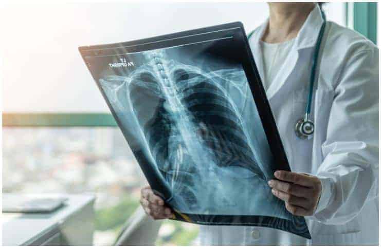
To check for any fluid accumulation or presence of any tumor and to detect the condition, your healthcare provider may do a CT scan (computerized tomography), as well as he may request for chest X-ray (uses small amounts of radiation to produce pictures of the organs).
Treatment
Treatment may include chest physical therapy, drug therapy, and sometimes surgery. However, the collapsed lung commonly expands once the cause has been corrected.
Pneumothorax
It is a collection of free air in the chest outside the lung, which causes the lung to collapse. Women are more likely to be affected around the age of 30 years and men in their early 20s.
Types
Primary spontaneous pneumothorax (PSP) – it usually occurs in healthy people. It is not caused by an injury, like – a rib fracture. Also, due to the rupture of small subpleural blebs, PSP commonly occurs in tall, thin young men.
Tension pneumothorax (TP) – it is usually caused by a lung laceration that allows air to escape into the pleural space. TP is a life-threatening condition that is more likely to occur in patients on mechanical ventilation.
Secondary pneumothorax (SP) – it occurs in the presence of existing lung disease as well as a cyst in chronic obstructive pulmonary disease or a rupture of a congenital bulla. Because of acute respiratory failure, SP usually requires admission to the intensive care unit.
READ MORE: Contact Dermatitis vs Scabies – Differences
Symptoms
Common symptoms may include:
- fatigue;
- chest pain which typically has a sudden onset;
- cough;
- rapid breathing;
- rapid heart rate;
- shortness of breath;
- a sharp pain that may lead to feelings of tightness in the chest.
Complications
Complications may include:
- respiratory failure;
- low blood oxygen;
- cardiac arrest.
Causes
Causes may include:
- certain lung diseases;
- drug abuse;
- cigarette smoking;
- trauma to the chest cavity.
Risk Factors
Risk factors include:
- if you already had this condition, you have an increased risk of another pneumothorax, commonly within two years of the first;
- if you need mechanical ventilation to assist your breathing, you are at higher risk of the condition;
- having an underlying lung disease, like – COPD;
- if you have between 20 and 40 years, particularly if you are underweight and very tall;
- if you smoke tobacco;
- if you are a man.
READ MORE: Myocarditis vs Endocarditis
Diagnosis
A chest x-ray shows the air pocket and the collapsed lung outlined by the thin inner pleural layer. Also, a physical examination can confirm the diagnosis if the pneumothorax is large.
Treatment
The treatment depends on the size of the clinical symptomatology, the pneumothorax, and the frequency of the occurrence.
In tension pneumothorax, the air needs to be removed from the pleural space (the thin fluid-filled space between the two pulmonary pleurae). This is typically done by inserting a long needle in between the ribs into the pleural space.
If the sufferer is not mechanically ventilated and the pneumothorax is small, his condition can be observed.
READ MORE: Smallpox vs Chickenpox
Atelectasis vs Pneumothorax (Collapsed Lung) – Differences
Atelectasis is the incomplete expansion of a lung or the airlessness or collapse of a lung that had once been expanded. It is one of the most common breathing complications after medical surgery.
A pneumothorax can be caused by certain medical procedures, a penetrating or blunt chest injury, or damage from underlying lung disease. The symptoms can vary from mild to life-threatening, and they include:
- loss of consciousness;
- shortness of breath;
- dizziness;
- chest pain, which may be more severe on one side of the chest;
- confusion;
- sharp pain when inhaling;
- rapid breathing;
- a pressure in the chest which gets worse over time;
- increased heart rate;
- blue discoloration of the skin or lips.
Note – sufferers must receive immediate emergency medical care in case breathing becomes increasingly difficult.
READ THIS NEXT: Dyslipidemia vs Hyperlipidemia
References

https://www.ncbi.nlm.nih.gov/pubmed/14104855
https://study.com/academy/lesson/what-is-atelectasis.html
https://www.sciencedirect.com/topics/medicine-and-dentistry/pneumothorax